")
")
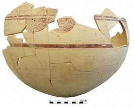
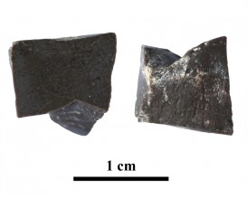
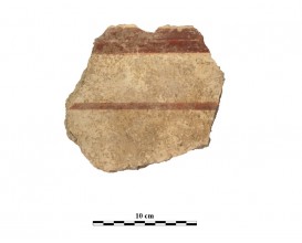
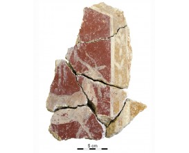
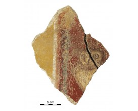
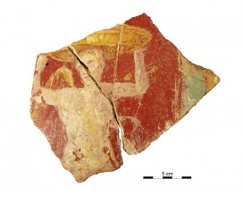
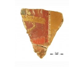
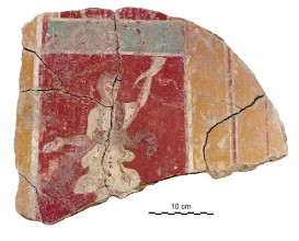
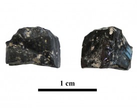
Wall fragment decorated with a quiver (Cástulo, Linares, Spain)
Dimensions
: 12.5 Centimeters
: 14 Centimeters
Materials
Painting
Temporal
: Roman
: Late 1st ct-2nd ct AD
Spatial
: Cástulo
: Linares, Jaén, Spain
: WGS84
Copyrights
Creative Commons - Attribution, Non-Commercial, No Derivatives (BY-NC-ND)
References
Digital Resources
-
 Conjunto arqueológico de Cástulo-Forum MMX
 Creative Commons - Attribution, Non-Commercial, No Derivatives (BY-NC-ND)
Arquiberlab
http://creativecommons.org/licenses/by-nc-nd/3.0/ -
 Conjunto arqueológico de Cástulo-Forum MMX
 Creative Commons - Attribution, Non-Commercial, No Derivatives (BY-NC-ND)
Arquiberlab
http://creativecommons.org/licenses/by-nc-nd/3.0/ -
 Conjunto arqueológico de Cástulo-Forum MMX
 Creative Commons - Attribution, Non-Commercial, No Derivatives (BY-NC-ND)
Arquiberlab
http://creativecommons.org/licenses/by-nc-nd/3.0/ -
 Instituto Univesitario de investigación en Arqueología Ibérica. Universidad de Jaén
 Creative Commons - Attribution, Non-Commercial, No Derivatives (BY-NC-ND)
Arquiberlab
http://creativecommons.org/licenses/by-nc-nd/3.0/ -
 Instituto Univesitario de investigación en Arqueología Ibérica. Universidad de Jaén
 Creative Commons - Attribution, Non-Commercial, No Derivatives (BY-NC-ND)
Arquiberlab
http://creativecommons.org/licenses/by-nc-nd/3.0/ -
 Instituto Univesitario de investigación en Arqueología Ibérica. Universidad de Jaén
 Creative Commons - Attribution, Non-Commercial, No Derivatives (BY-NC-ND)
Arquiberlab
http://creativecommons.org/licenses/by-nc-nd/3.0/ -
 Instituto Univesitario de investigación en Arqueología Ibérica. Universidad de Jaén
 Creative Commons - Attribution, Non-Commercial, No Derivatives (BY-NC-ND)
Arquiberlab
http://creativecommons.org/licenses/by-nc-nd/3.0/ -
 Instituto Univesitario de investigación en Arqueología Ibérica. Universidad de Jaén
 Creative Commons - Attribution, Non-Commercial, No Derivatives (BY-NC-ND)
Arquiberlab
http://creativecommons.org/licenses/by-nc-nd/3.0/ - Instituto Univesitario de investigación en Arqueología Ibérica. Universidad de Jaén
Creative Commons - Attribution, Non-Commercial, No Derivatives (BY-NC-ND)
Arquiberlab
http://creativecommons.org/licenses/by-nc-nd/3.0/
Activities
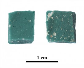
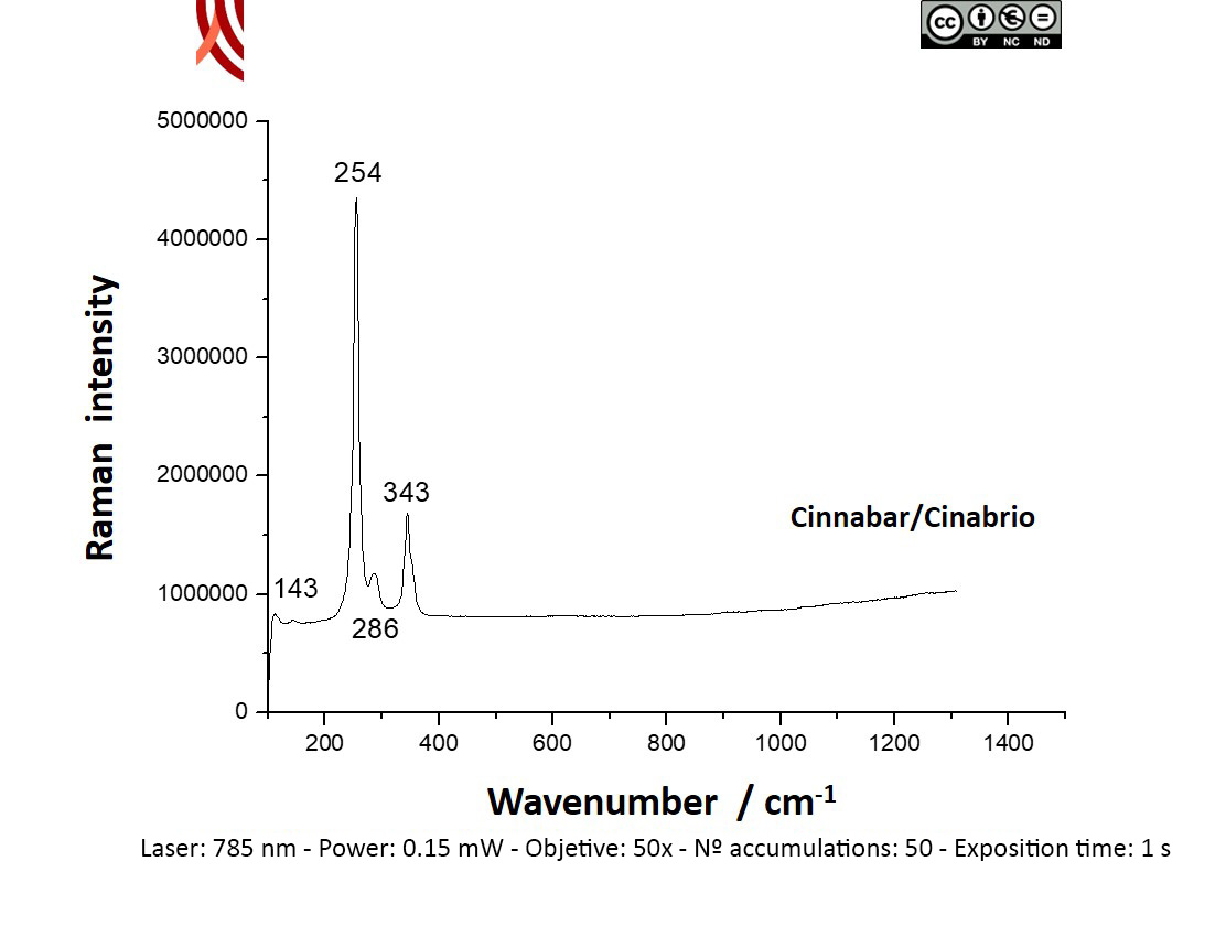
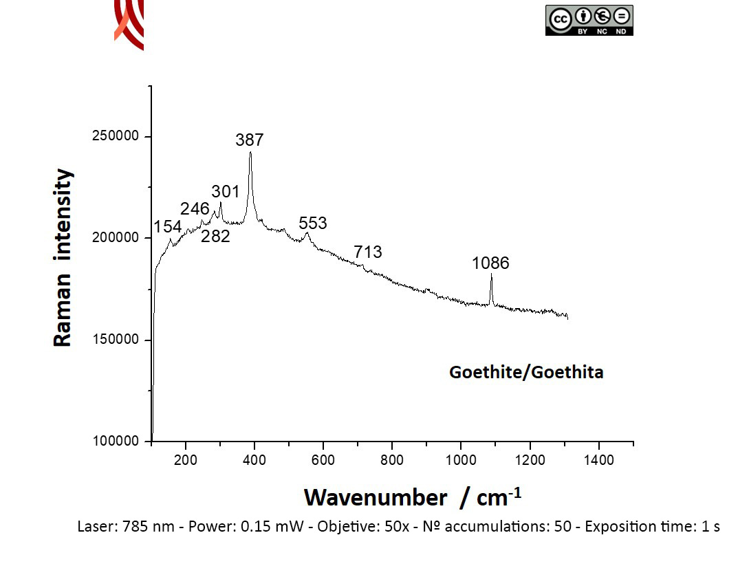
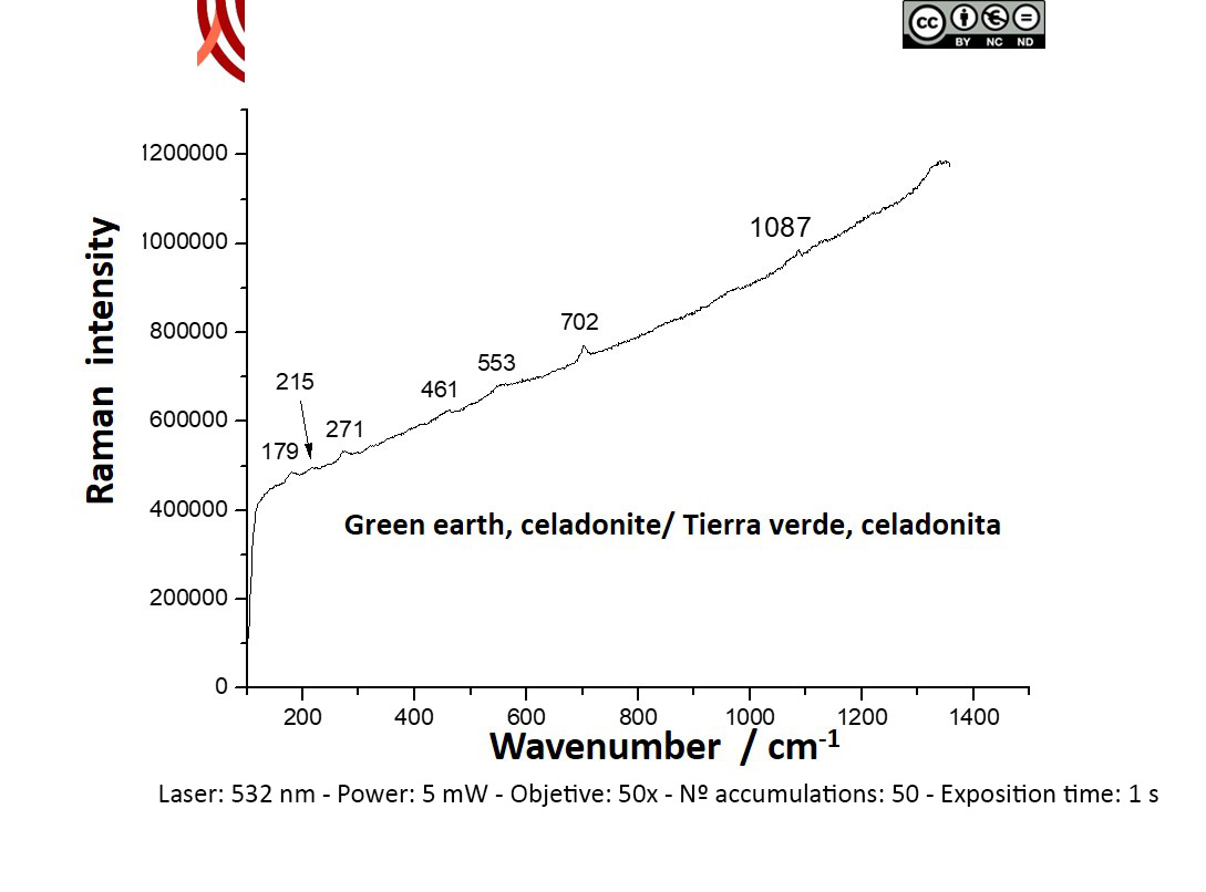
Archaeometric analysis Physical-chemical analysis Wall painting. Decoration analysis.
| |
Raman Microscopy Mineral analysis Non destructive. Surface cleaning. Sample pretreatment is not required. Direct measurement. Micro-Raman Spectroscopy (MRS) Equipment 1: Renishaw ‘in via’ Reflex Spectrometer coupled with a confocal Leica DM LM microscope (CICT, University of Jaén), equipped with a diode laser (785 nm, 300 mW), argon ion laser (514.5 nm, 25 mW) and a Peltier-cooled CCD detector, calibrated to the 520.5 cm-1 line of silicon.; ; Equipment 2: Renishaw ‘in via’ Qontor Spectrometer coupled with a confocal Leica DM LM microscope (SCAI, University of Málaga), equipped with a solid state laser (473 nm, 25 mW), a Nd:YAG laser (532 nm, 50 mW) and a diode laser (785 nm, 300 mW), and a Peltier-cooled CCD detector, calibrated to the 520.5 cm-1 line of silicon. | |
X-Ray Fluorescence Elemental analysis Non destructive. Surface cleaning. Sample pretreatment is not required. Direct measurement. Energy dispersive X-ray fluorescence (EDXRF) An energy dispersive X-ray microfluorescence spectrometer (M4 Tornado, Bruker) (CICT, University of Jaen). This spectrometer is equipped with a microfocus X-ray tube with an Rh anode, a polycapillary lens for X-ray focussing, and a 30-mm2 energy dispersive detector (SDD). The sample chamber incorporates an XYZ motorized stage for sample positioning. A high resolution microscope is used to position the sample on the desired distance from the polycapillary. To increase the sensitivity of the low Z elements, the sample chamber can be brought under vacuum. For the analysis of the samples, a spot size of 25 μm was chosen at an operating X-ray tube voltage of 50 kV and intensity of 600 μA. The tube current was adapted for each sample in order to optimise the detection of X-rays. |