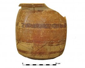
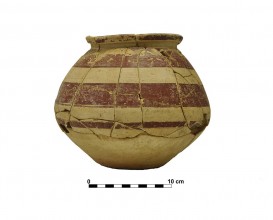
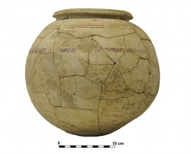
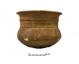
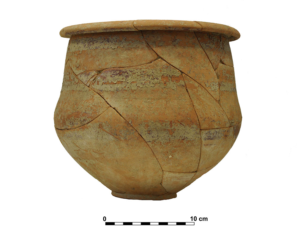
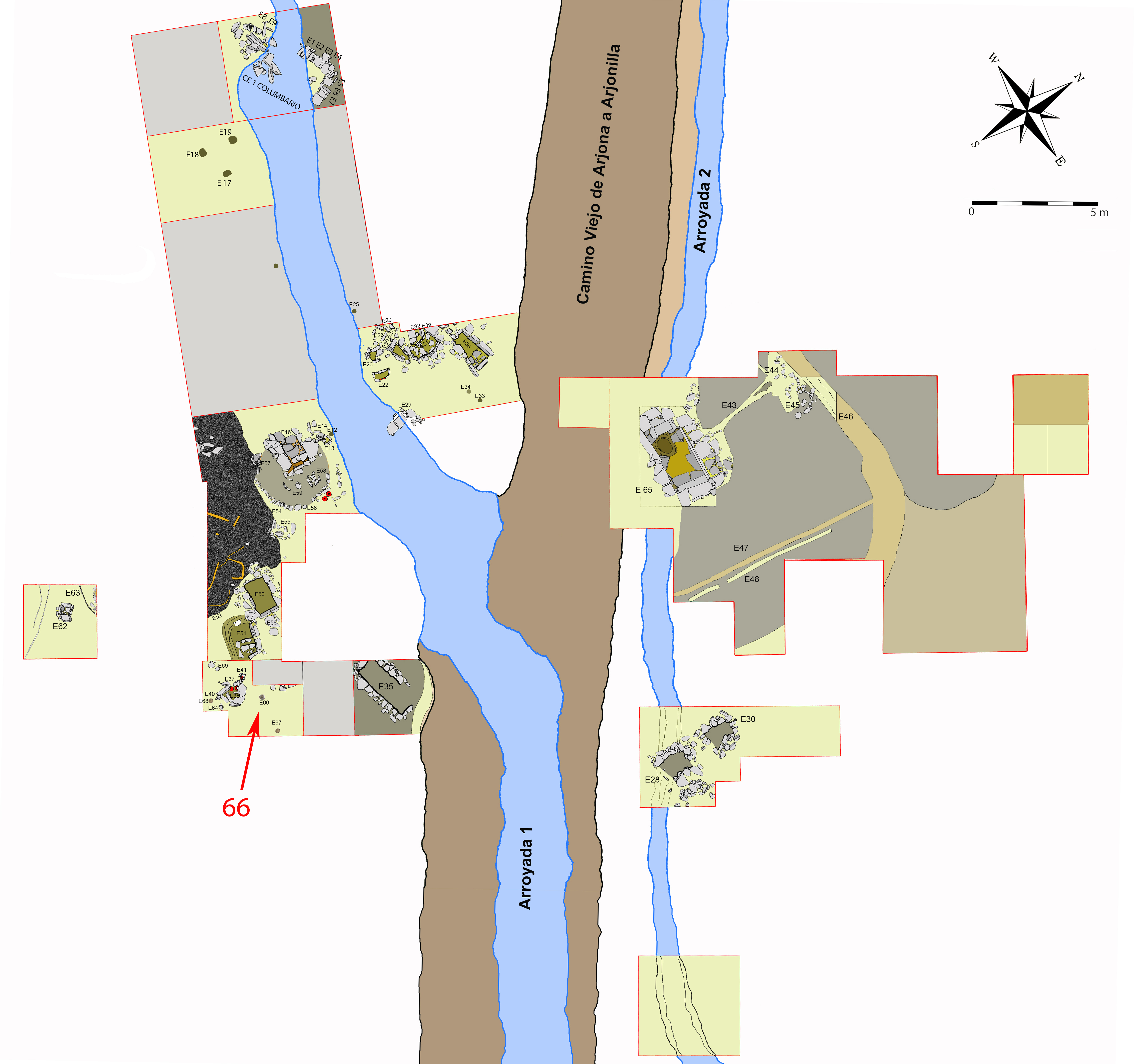
Ceramic vessel 5. Grave 66. Cemetery of Piquía
Dimensions
: 19 Centimeters
: 19 Centimeters
Materials
pottery
Temporal
: Iberian, Iberians
: 1st ct. BC
Spatial
: Cemetery of Piquía
: Arjona, Jaén, Spain
: WGS84
Copyrights
Creative Commons - Attribution, Non-Commercial, No Derivatives (BY-NC-ND)
References
Ruiz, A., Molinos, M., Gómez, F y Lechuga M. A. (2015): “La cámara de Piquía, Arjona”, en A. Ruiz y M. Molinos (coord.): Jaén, tierra ibera. 40 años de investigación y transferencia. Universidad de Jaén. 357-374.
Digital Resources
-
Creative Commons - Attribution, Non-Commercial, No Derivatives (BY-NC-ND)
Arquiberlab
http://creativecommons.org/licenses/by-nc-nd/3.0/ -
Creative Commons - Attribution, Non-Commercial, No Derivatives (BY-NC-ND)
Arquiberlab
http://creativecommons.org/licenses/by-nc-nd/3.0/ -
Creative Commons - Attribution, Non-Commercial, No Derivatives (BY-NC-ND)
Arquiberlab
http://creativecommons.org/licenses/by-nc-nd/3.0/ -
Creative Commons - Attribution, Non-Commercial, No Derivatives (BY-NC-ND)
Arquiberlab
http://creativecommons.org/licenses/by-nc-nd/3.0/ -
Creative Commons - Attribution, Non-Commercial, No Derivatives (BY-NC-ND)
Arquiberlab
http://creativecommons.org/licenses/by-nc-nd/3.0/
Activities
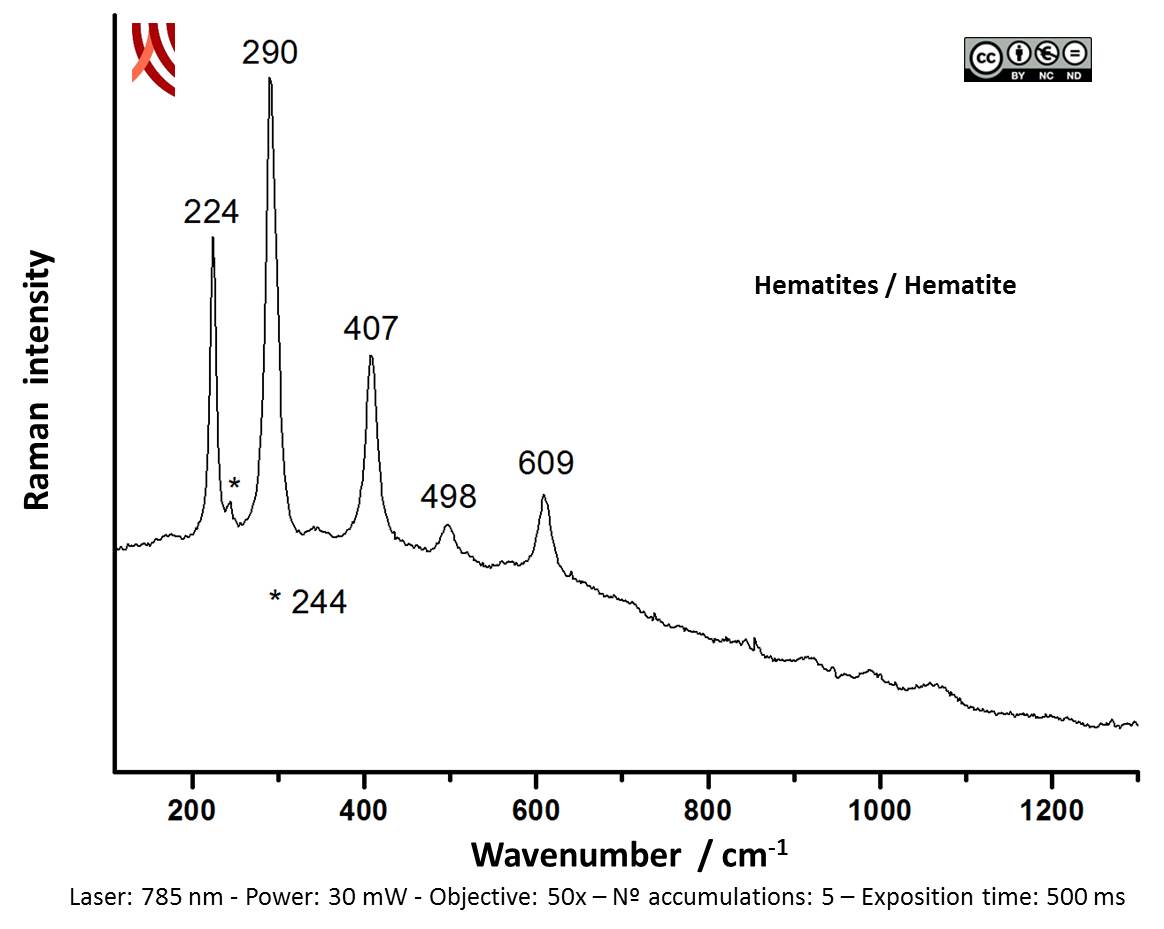
Archaeometric analysis Physical-chemical analysis Ceramic. Analysis of decoration
| |
Raman Microscopy Mineral analisis of the red decoration Non destructive. Surface cleaning. Sample pretreatment is not required. Direct measurement. Micro-Raman Spectroscopy (MRS) Portable equipment: BWS445-785S innoRam™ Raman spectrometer (B%26WTEK, Inc., Newark, USA) with a 785 nm excitation laser (maximum power of 300 mW) and a 4.5 cm-1 spectral resolution. The Raman microprobe can be mounted on a tripod with motorized XYZ axis (MICROBEAM S.A, Barcelona, Spain) or on a microscope sampling stage (B%26WTEK, Inc., Newark, USA). | |
X-Ray Fluorescence Elemental analysis of the red decoration Non destructive. Surface cleaning. Sample pretreatment is not required. Direct measurement. Energy dispersive X- ray fluorescence (EDXRF) EDAX (model Eagle III) fluorescence spectrometer (CITI, University of Seville). This spectrometer is equipped with a microfocus X-ray tube with an Rh anode, a polycapillary lens for X-ray focussing, and an 80 mm2 energy dispersive Si-(Li) detector. The sample chamber incorporates an XYZ motorized stage for sample positioning. A high resolution microscope is used to position the sample on the desired distance from the polycapillary. To increase the sensitivity of the low Z elements, the sample chamber can be brought under vacuum. For the analysis of the samples, a spot size of 300 μm was chosen at an operating X-ray tube voltage of 40 kV. The tube current was adapted for each sample in order to optimise the detection of X-rays |