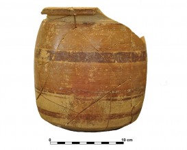
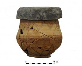
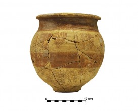
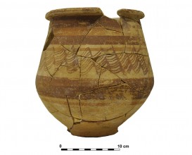
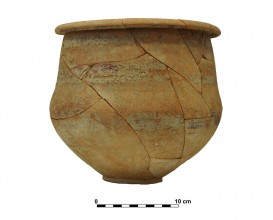
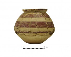
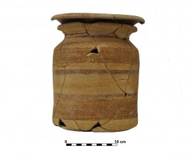
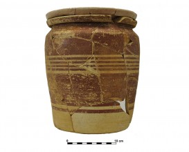
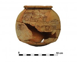
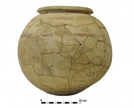
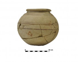
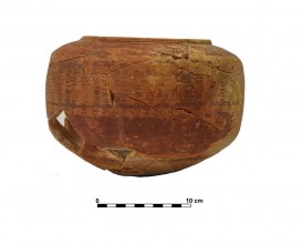
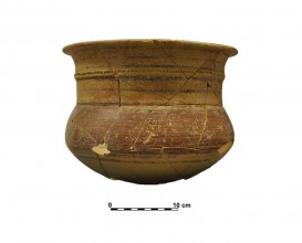
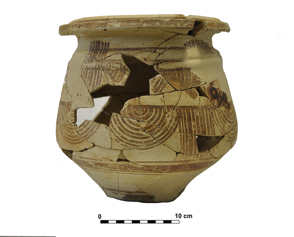
Ceramic vessel 12. Grave 65. Cemetery of Piquía
Dimensions
: 24 Centimeters
: 25 Centimeters
Materials
pottery
Temporal
: Iberian, Iberians
: 1st ct. BC
Spatial
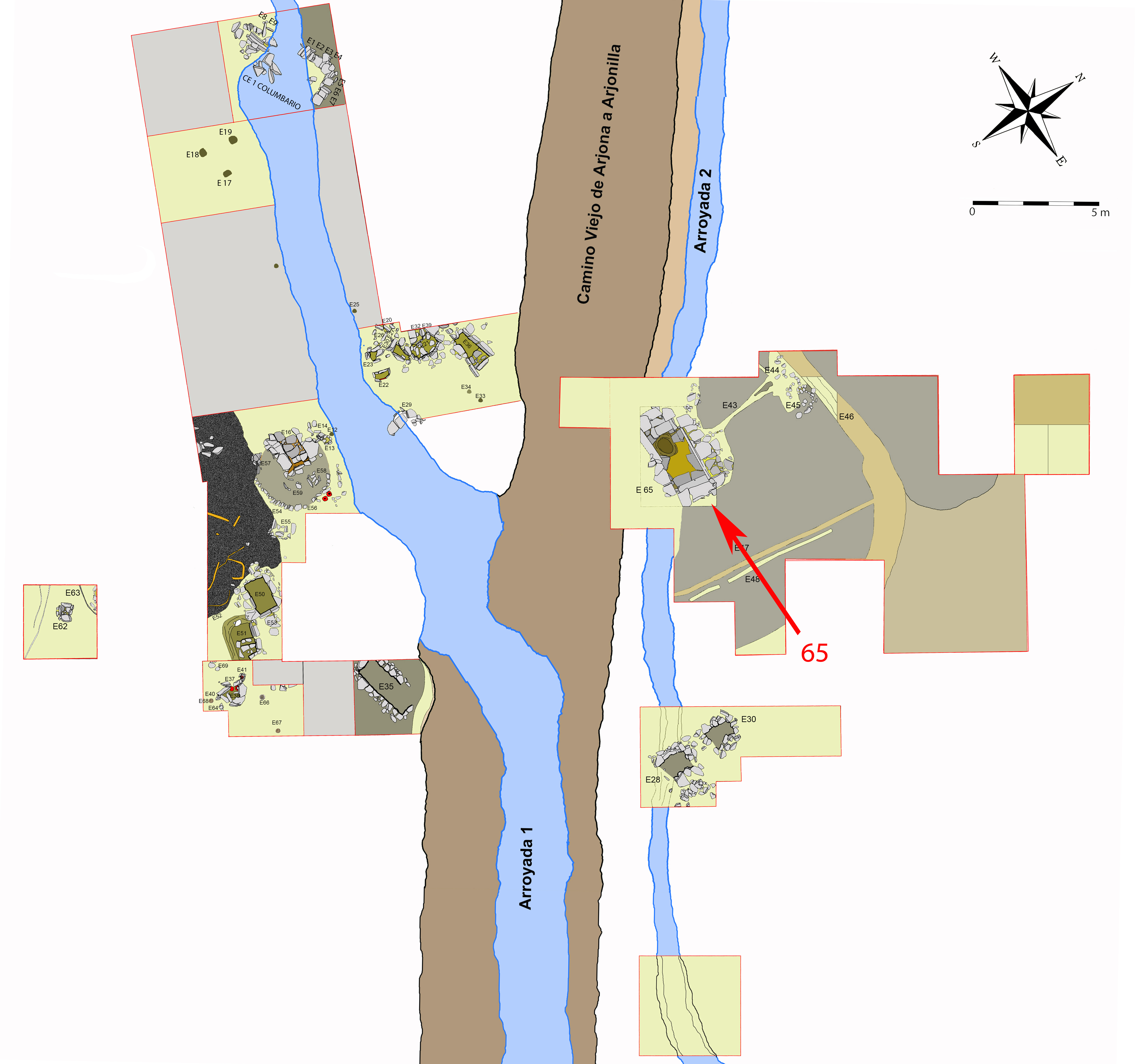
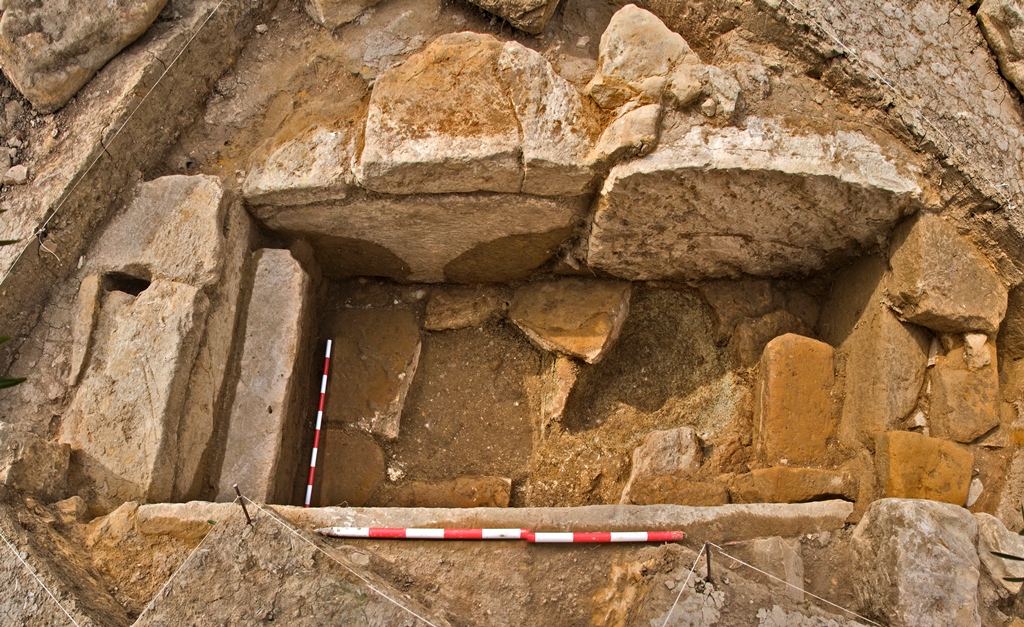
: Cemetery of Piquía
: Arjona, Jaén, Spain
: WGS84
Copyrights
Creative Commons - Attribution, Non-Commercial, No Derivatives (BY-NC-ND)
References
Ruiz, A., Molinos, M., Gómez, F y Lechuga M. A. (2015): “La cámara de Piquía, Arjona”, en A. Ruiz y M. Molinos (coord.): Jaén, tierra ibera. 40 años de investigación y transferencia. Universidad de Jaén. 357-374.
Digital Resources
-
Creative Commons - Attribution, Non-Commercial, No Derivatives (BY-NC-ND)
Arquiberlab
http://creativecommons.org/licenses/by-nc-nd/3.0/ -
Creative Commons - Attribution, Non-Commercial, No Derivatives (BY-NC-ND)
Arquiberlab
http://creativecommons.org/licenses/by-nc-nd/3.0/ -
Creative Commons - Attribution, Non-Commercial, No Derivatives (BY-NC-ND)
Arquiberlab
http://creativecommons.org/licenses/by-nc-nd/3.0/ -
Creative Commons - Attribution, Non-Commercial, No Derivatives (BY-NC-ND)
Arquiberlab
http://creativecommons.org/licenses/by-nc-nd/3.0/ -
Creative Commons - Attribution, Non-Commercial, No Derivatives (BY-NC-ND)
Arquiberlab
http://creativecommons.org/licenses/by-nc-nd/3.0/ -
Creative Commons - Attribution, Non-Commercial, No Derivatives (BY-NC-ND)
Arquiberlab
http://creativecommons.org/licenses/by-nc-nd/3.0/ -
Creative Commons - Attribution, Non-Commercial, No Derivatives (BY-NC-ND)
Arquiberlab
http://creativecommons.org/licenses/by-nc-nd/3.0/
Activities
Archaeometric analysis Physical-chemical analysis Ceramic. Analysis of decoration
| |
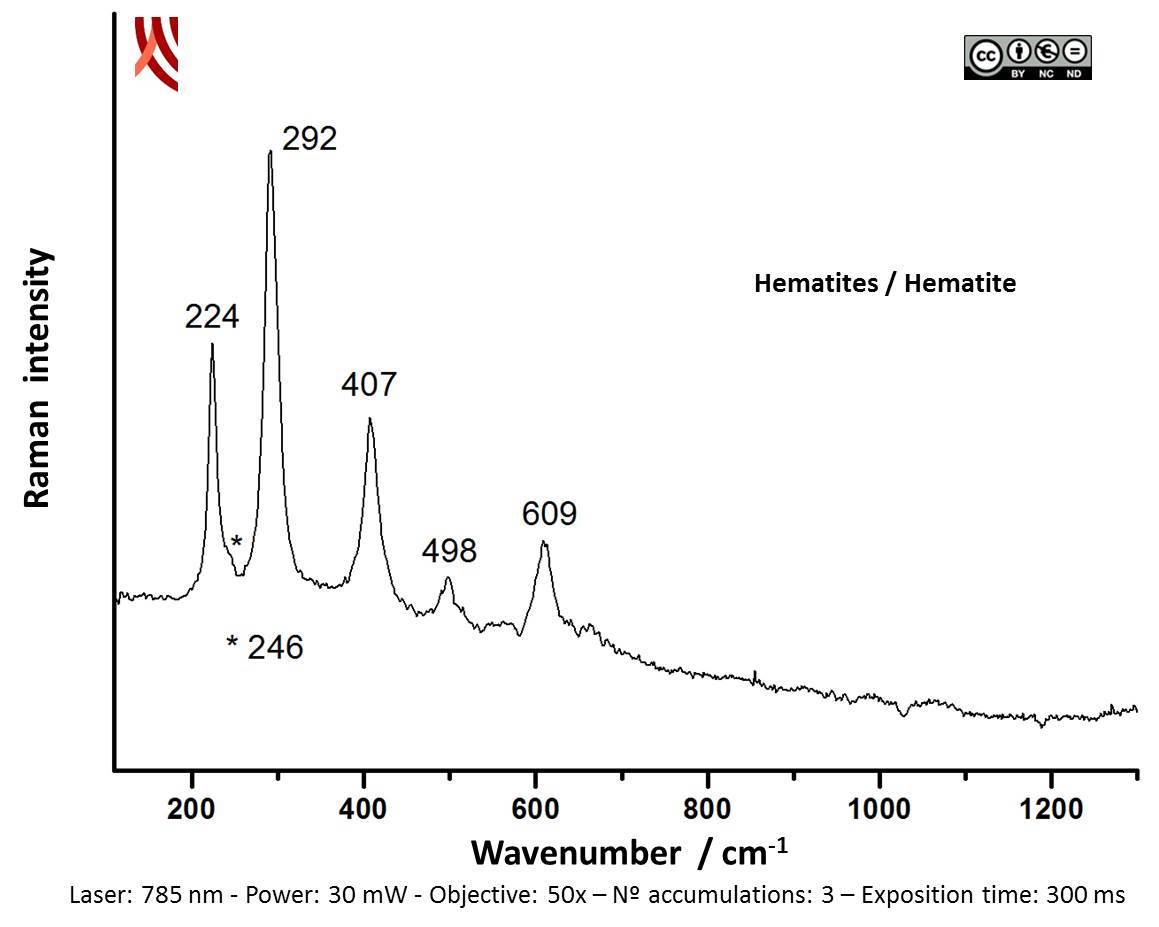
Raman Microscopy Mineral analysis of the red decoration Non destructive. Surface cleaning. Sample pretreatment is not required. Direct measurement. Micro-Raman Spectroscopy (MRS) Portable equipment: BWS445-785S innoRam™ Raman spectrometer (B%26WTEK, Inc., Newark, USA) with a 785 nm excitation laser (maximum power of 300 mW) and a 4.5 cm-1 spectral resolution. The Raman microprobe can be mounted on a tripod with motorized XYZ axis (MICROBEAM S.A, Barcelona, Spain) or on a microscope sampling stage (B%26WTEK, Inc., Newark, USA). | |
X-Ray Fluorescence Elemental analysis of the red decoration Non destructive. Surface cleaning. Sample pretreatment is not required. Direct measurement. Energy dispersive X- ray fluorescence (EDXRF); Espectrómetro de fluorescencia de rayos X de energía dispersiva EDAX (modelo Eagle III)( CITI, Universidad de Sevilla). Está equipado con un tubo de rayos X microfocus con un ánodo de Rh, una lente policapilar para enfoque de rayos X y un detector de energía dispersiva Si-(Li) de 80mm2. La cámara de medida incorpora una bandeja motorizada para el posicionamiento de la muestra. Microscopio de alta resolución para posicionar la muestra a la distancia adecuada. Cámara con bomba de vacío para elementos de bajo número atómico (Z). Para un tamaño del área de medida de 300 μm se selecciona en el tubo de rayos X un voltaje de 40kV. La intensidad de corriente del tubo se adapta a cada muestra para optimizar la detección de los rayos X. |