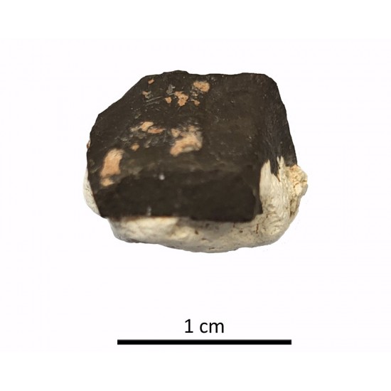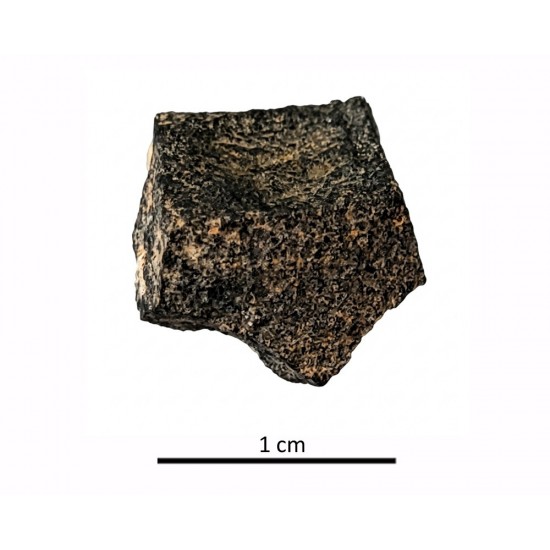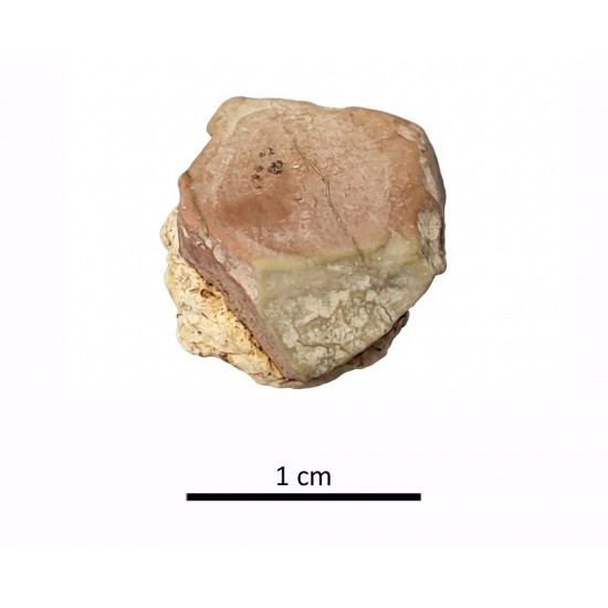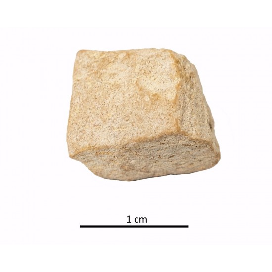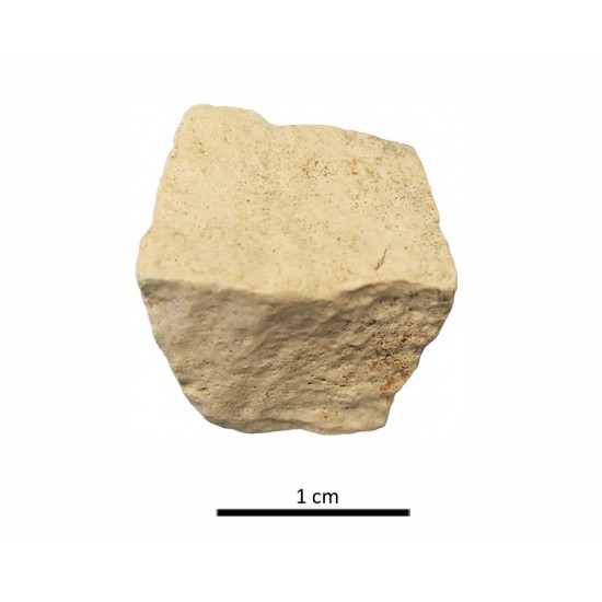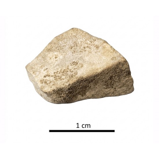
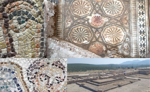



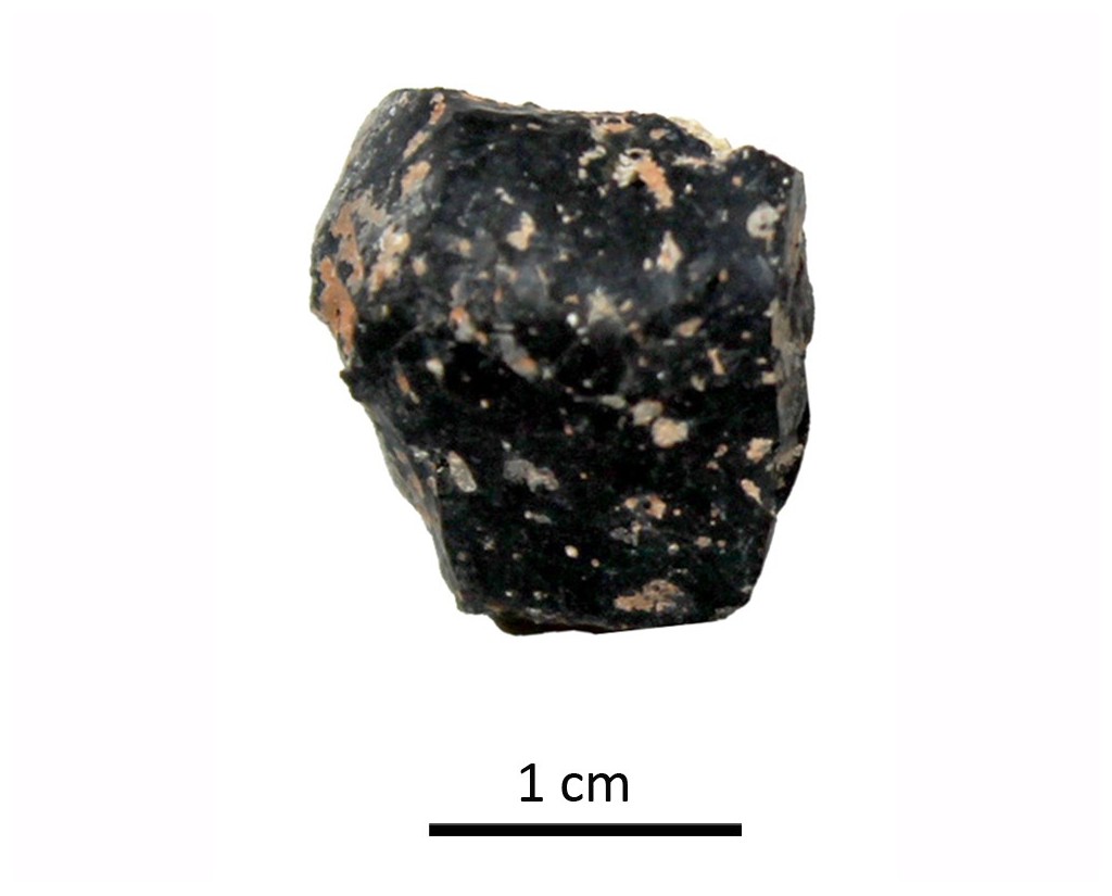

Tessera 1. Villa Cerrillo del Cuco (Vilches, Jaén, Spain)
Dimensions
: 1 Centimeters
: 1 Centimeters
: 1 Centimeters
Materials
glass paste
Temporal
: Roman
: 5th century AD
Spatial
- : Villa Cerrillo del Cuco
- : Vilches, Jaén, Spain
- : WGS84
Copyrights
Creative Commons - Attribution, Non-Commercial, No Derivatives (BY-NC-ND)
References
Molinos, M., Ruiz, A y Unghetti, C. (1981): Excavaciones arqueológicas en la villa romana del Cerrillo del Cuco (Santagón, Vilches, Jaén). Actas del 1 Congreso Andaluz de Estudios Clásicos, pp. 306-312.
Nueva Referencia
<
Blázquez, J. Mª, López, G., San Nicolás, Mª P., Neira, Mª L. (1987): Mosaico romano de Vilches (Jaén). Archivo Español de Arqueología, 60: 275-279.
Digital Resources
-

Creative Commons - Attribution, Non-Commercial, No Derivatives (BY-NC-ND)
Arquiberlab
http://creativecommons.org/licenses/by-nc-nd/3.0/
[ver más...] [ver menos] -

Creative Commons - Attribution, Non-Commercial, No Derivatives (BY-NC-ND)
Arquiberlab
http://creativecommons.org/licenses/by-nc-nd/3.0/
[ver más...] [ver menos] -

Creative Commons - Attribution, Non-Commercial, No Derivatives (BY-NC-ND)
Arquiberlab
http://creativecommons.org/licenses/by-nc-nd/3.0/
[ver más...] [ver menos] -
 IUIAI-UJA
Creative Commons - Attribution, Non-Commercial, No Derivatives (BY-NC-ND)
Arquiberlab
http://creativecommons.org/licenses/by-nc-nd/3.0/
[ver más...] [ver menos] -
 IUIAI-UJA
Creative Commons - Attribution, Non-Commercial, No Derivatives (BY-NC-ND)
Arquiberlab
http://creativecommons.org/licenses/by-nc-nd/3.0/
[ver más...] [ver menos] -
 IUIAI-UJA
Creative Commons - Attribution, Non-Commercial, No Derivatives (BY-NC-ND)
Arquiberlab
http://creativecommons.org/licenses/by-nc-nd/3.0/
[ver más...] [ver menos] -
IUIAI-UJA
Creative Commons - Attribution, Non-Commercial, No Derivatives (BY-NC-ND)
Arquiberlab
http://creativecommons.org/licenses/by-nc-nd/3.0/
[ver más...] [ver menos]
Activities
University Research Center for Iberian Archaeology |
|
Raman Microscopy Mineralogical analysis The tessera was sectioned to expose fresh, unaltered cross-section Renishaw ‘in via’ Qontor Spectrometer coupled with a confocal Leica DM LM microscope (CICT, University of Jaén), equipped with a diode laser (785 nm, 300 mW), DPSS laser (532 nm, 50 mW) and a Peltier-cooled CCD detector, calibrated to the 520.5 cm-1 line of silicon. |
|
X-Ray Fluorescence Elemental analysis The tessera was sectioned to expose fresh, unaltered cross-section EDAX (model Eagle III) fluorescence spectrometer (CITI, University of Seville). This spectrometer is equipped with a microfocus X-ray tube with an Rh anode, a polycapillary lens for X-ray focussing, and an 80 mm2 energy dispersive Si-(Li) detector. The sample chamber incorporates an XYZ motorized stage for sample positioning. A high resolution microscope is used to position the sample on the desired distance from the polycapillary. To increase the sensitivity of the low Z elements, the sample chamber can be brought under vacuum. For the analysis of the samples, a spot size of 300 μm was chosen at an operating X-ray tube voltage of 40 kV. The tube current was adapted for each sample in order to optimise the detection of X-rays. |