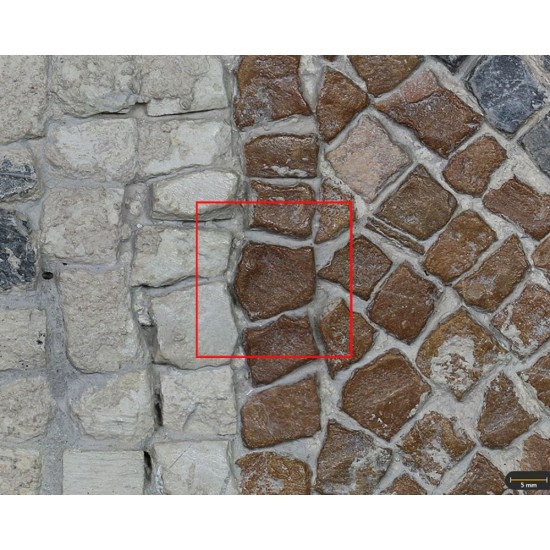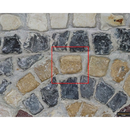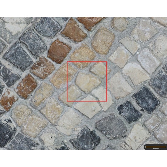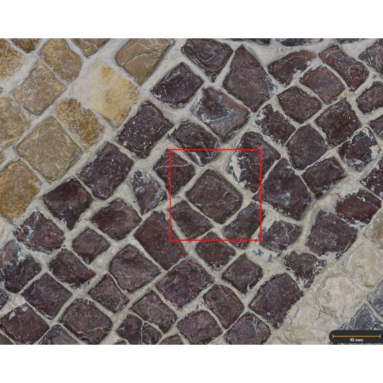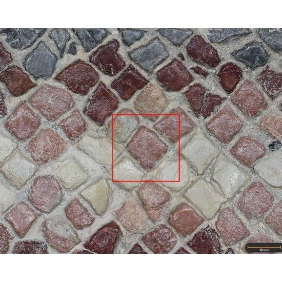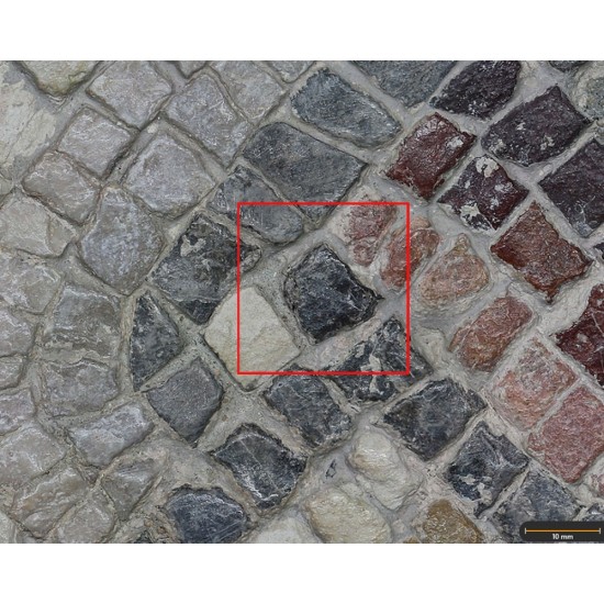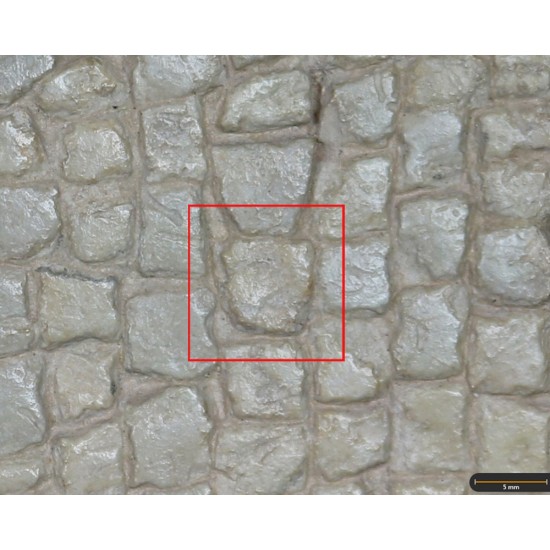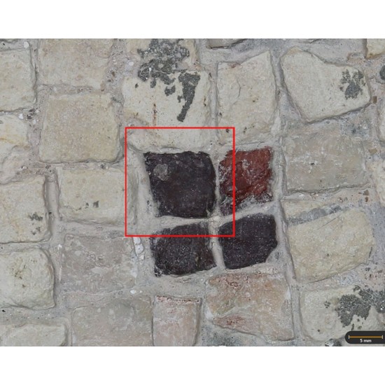
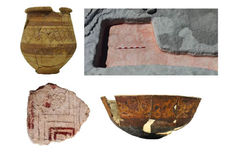
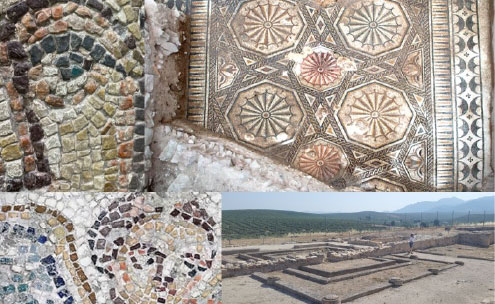



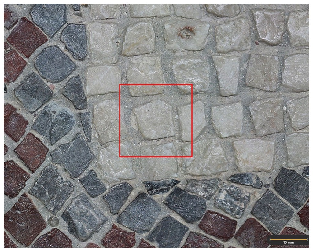

Tessera 20. Tethys mosaic, Villa of Bruñel (Quesada, Jaén, Spain)
Dimensions
: 1 Centimeters
: 1 Centimeters
Materials
Stone
Temporal
: Roman
: 3rd-4th century AD
Spatial
- : Villa de Bruñel
- : Quesada, Jaén, Spain
- : WGS84
Copyrights
Creative Commons - Attribution, Non-Commercial, No Derivatives (BY-NC-ND)
References
Digital Resources
-

Creative Commons - Attribution, Non-Commercial, No Derivatives (BY-NC-ND)
Arquiberlab
http://creativecommons.org/licenses/by-nc-nd/3.0/
[ver más...] [ver menos] -

Creative Commons - Attribution, Non-Commercial, No Derivatives (BY-NC-ND)
Arquiberlab
http://creativecommons.org/licenses/by-nc-nd/3.0/
[ver más...] [ver menos] -

Creative Commons - Attribution, Non-Commercial, No Derivatives (BY-NC-ND)
Arquiberlab
http://creativecommons.org/licenses/by-nc-nd/3.0/
[ver más...] [ver menos] -
 IUIAI-UJA
Creative Commons - Attribution, Non-Commercial, No Derivatives (BY-NC-ND)
Arquiberlab
http://creativecommons.org/licenses/by-nc-nd/3.0/
[ver más...] [ver menos] -
IUIAI-UJA
Creative Commons - Attribution, Non-Commercial, No Derivatives (BY-NC-ND)
Arquiberlab
http://creativecommons.org/licenses/by-nc-nd/3.0/
[ver más...] [ver menos]
Activities
University Research Center for Iberian Archaeology |
|
Raman Microscopy Mineralogical analysis Non destructive. Surface cleaning. Sample pretreatment is not required. Direct measurement. Portable Raman spectrometer (model BWS445-785S innoRam) equipped with a CCD detector, a handheld fiber-optic probe, an automated MicroBeam tripod, a benchtop stand for small samples, and a BAC151A camera (University Research Institute for Iberian Archaeology, University of Jaén). A 785 nm diode laser (300 mW) was used, with a spectral resolution better than 4.5 cm-1 |
|
X-Ray Fluorescence Elemental analysis Non destructive. Surface cleaning. Sample pretreatment is not required. Direct measurement. Bruker Tracer III SD handheld XRF spectrometer (HERCULES Laboratory, University of Évora, Portugal). This instrument is equipped with a silicon drift 10 mm 2 SDD detector, and a Rh target delivering a polychromatic X-ray beam of 3×3 mm². Spectra were acquired with 40 kV and a current of 11 μA, for a life time of 30 s. This analytical method detects elements with an atomic number higher than 12. |