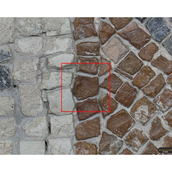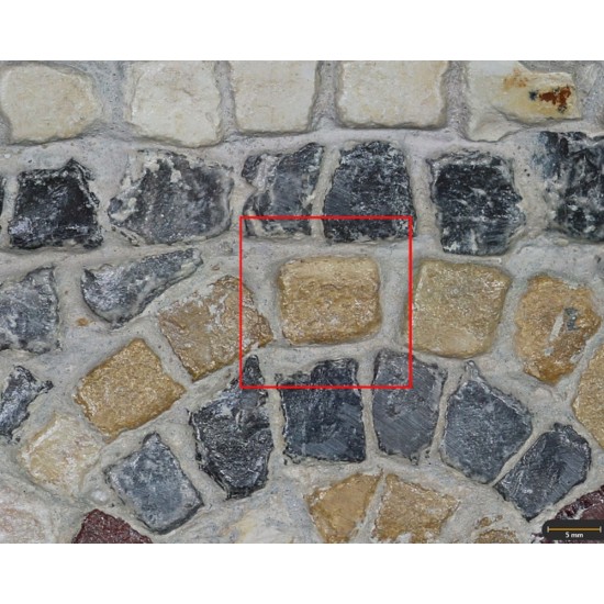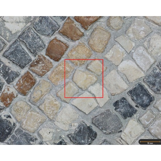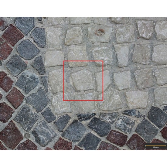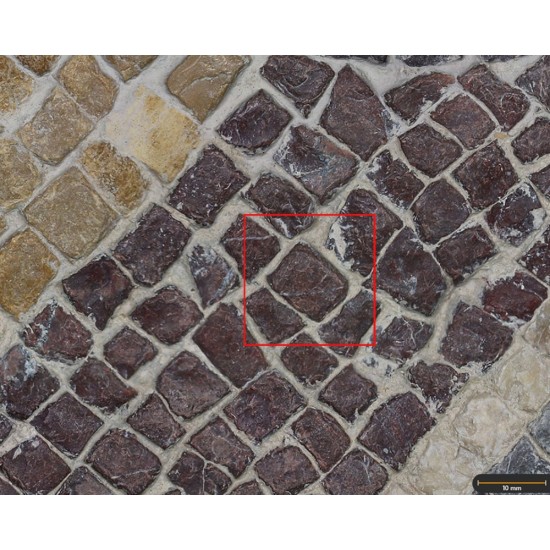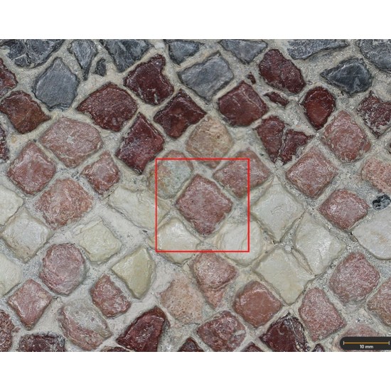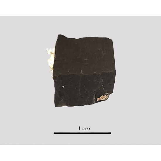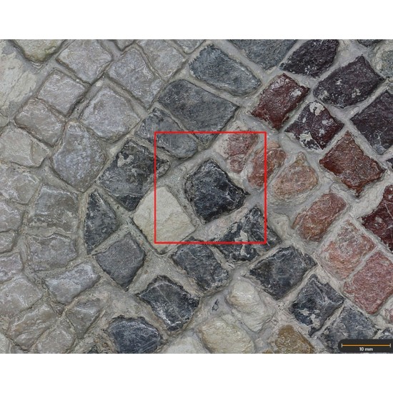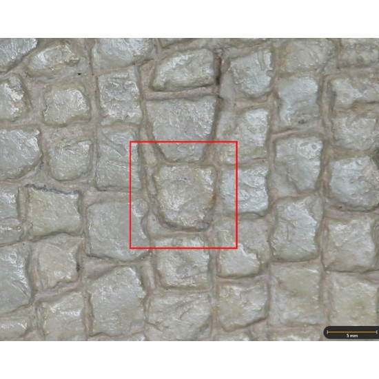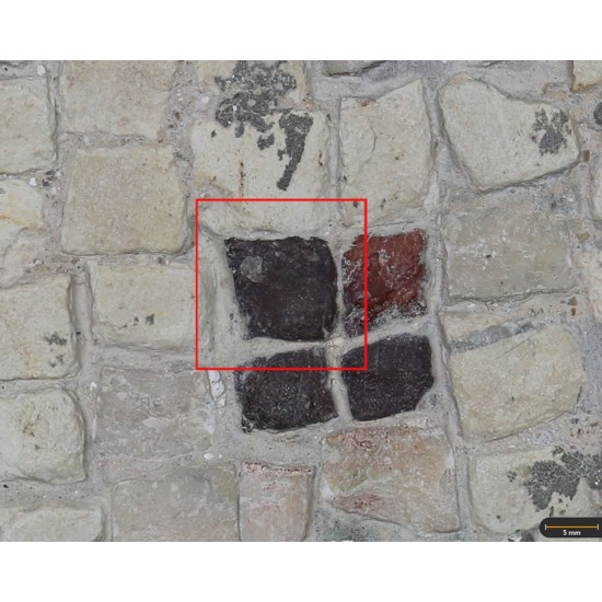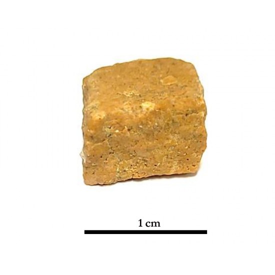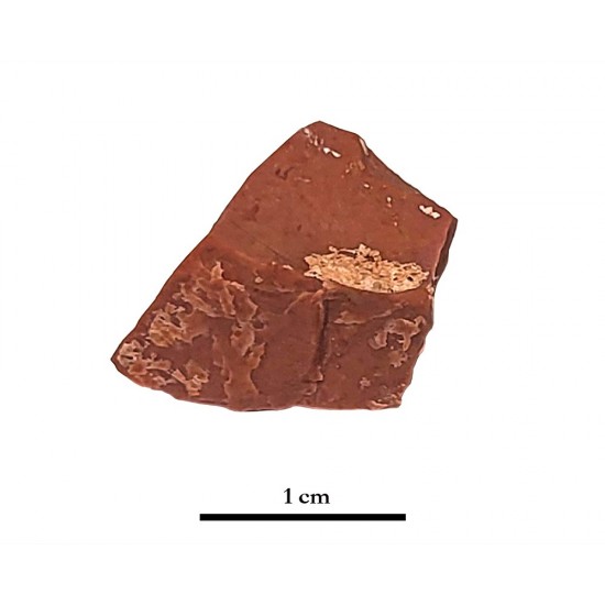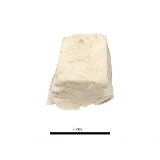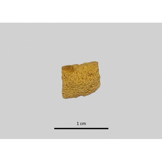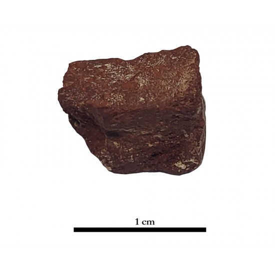
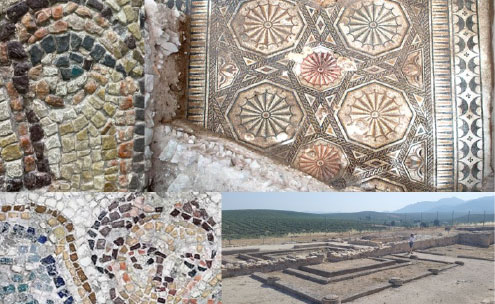



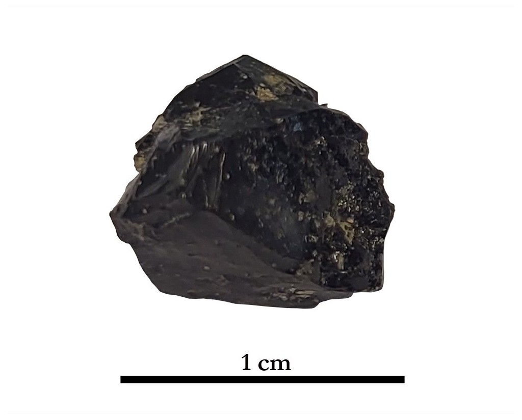

Tessera 1001-0. Villa de Bruñel (Quesada, Jaén, Spain)
The Raman spectrum provides further evidence for the presence of Fe- and Zn-bearing crystalline phases within the vitreous matrix. The bands at 532 and 683 cm⁻¹ are compatible respectively, of an Fe-bearing spinel and are particularly consistent with magnetite (Fe₃O₄), The relatively high Zn content (14.10 wt % ZnO) is consistent with the polymetallic Pb–Zn character of the Linares–La Carolina ores, where galena (PbS) and sphalerite (ZnS) are documented as associated ore minerals. However, the very low sulphur content of the tesserae (0.03 wt % SO₃) provides no evidence for the preservation of residual sphalerite. Instead, Zn was likely redistributed during the high-temperature smelting process and incorporated into the vitreous matrix and/or Zn–Fe-bearing crystalline phases. Thus, the Raman data are interpreted as indicating the presence of Fe- and/or Zn–Fe-bearing spinel phases formed during the metallurgical process, rather than residual sulphide minerals.
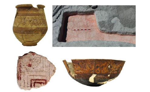
All of the above points to the conclusion that vitreous slag from galena smelting—materials commonly found in other mosaics of the Upper Guadalquivir region, such as those at El Altillo, Cerrillo del Cuco, and Los Amores—was reused in the creation of the mosaics at the Bruñel villa.
Dimensions
: 1 Centimeters
: 1 Centimeters
: 1 Centimeters
Materials
glass paste
Temporal
: Roman
: 3rd-4th century AD
Spatial
- : Villa de Bruñel
- : Quesada, Jaén, Spain
- : WGS84
Copyrights
Creative Commons - Attribution, Non-Commercial, No Derivatives (BY-NC-ND)
References
Digital Resources
-
 IUIAI-UJA
Creative Commons - Attribution, Non-Commercial, No Derivatives (BY-NC-ND)
Arquiberlab
http://creativecommons.org/licenses/by-nc-nd/3.0/
[ver más...] [ver menos] -
 IUIAI-UJA
Creative Commons - Attribution, Non-Commercial, No Derivatives (BY-NC-ND)
Arquiberlab
http://creativecommons.org/licenses/by-nc-nd/3.0/
[ver más...] [ver menos] -
 IUIAI-UJA
Creative Commons - Attribution, Non-Commercial, No Derivatives (BY-NC-ND)
Arquiberlab
http://creativecommons.org/licenses/by-nc-nd/3.0/
[ver más...] [ver menos] -
 CC-BY-NC
Creative Commons - Attribution, Non-Commercial, No Derivatives (BY-NC-ND)
Arquiberlab
http://creativecommons.org/licenses/by-nc-nd/3.0/
[ver más...] [ver menos] -
IUIAI-UJA
Creative Commons - Attribution, Non-Commercial, No Derivatives (BY-NC-ND)
Arquiberlab
http://creativecommons.org/licenses/by-nc-nd/3.0/
[ver más...] [ver menos]
Activities
University Research Center for Iberian Archaeology |
|
Raman Microscopy Mineralogical analysis The tessera was sectioned to expose fresh, unaltered cross-section Renishaw ‘in via’ Qontor Spectrometer coupled with a confocal Leica DM LM microscope (CICT, University of Jaén), equipped with a diode laser (785 nm, 300 mW), DPSS laser (532 nm, 50 mW) and a Peltier-cooled CCD detector, calibrated to the 520.5 cm-1 line of silicon. |
|
X-Ray Fluorescence Elemental analysis The tessera was sectioned to expose fresh, unaltered cross-section EDAX (model Eagle III) fluorescence spectrometer (CITI, University of Seville). This spectrometer is equipped with a microfocus X-ray tube with an Rh anode, a polycapillary lens for X-ray focussing, and an 80 mm2 energy dispersive Si-(Li) detector. The sample chamber incorporates an XYZ motorized stage for sample positioning. A high resolution microscope is used to position the sample on the desired distance from the polycapillary. To increase the sensitivity of the low Z elements, the sample chamber can be brought under vacuum. For the analysis of the samples, a spot size of 300 μm was chosen at an operating X-ray tube voltage of 40 kV. The tube current was adapted for each sample in order to optimise the detection of X-rays. |