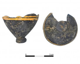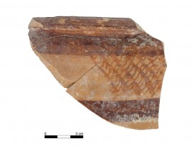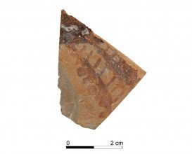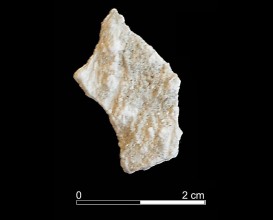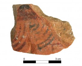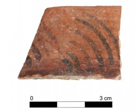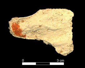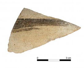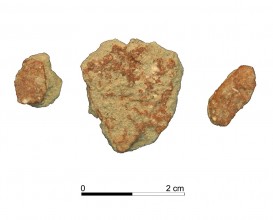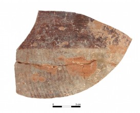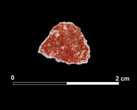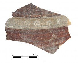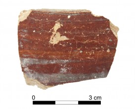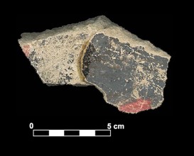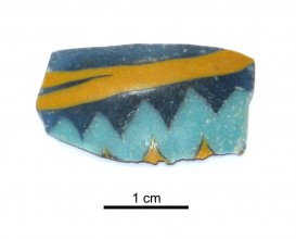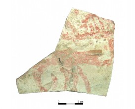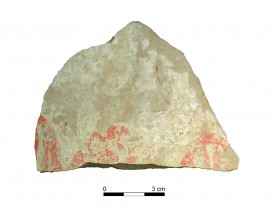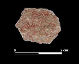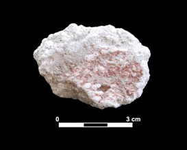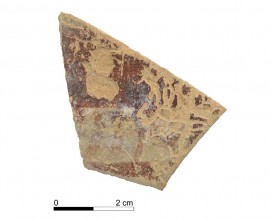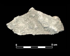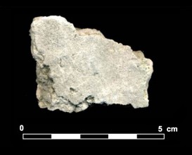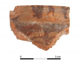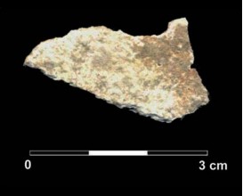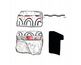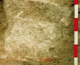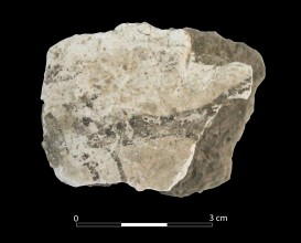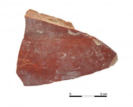



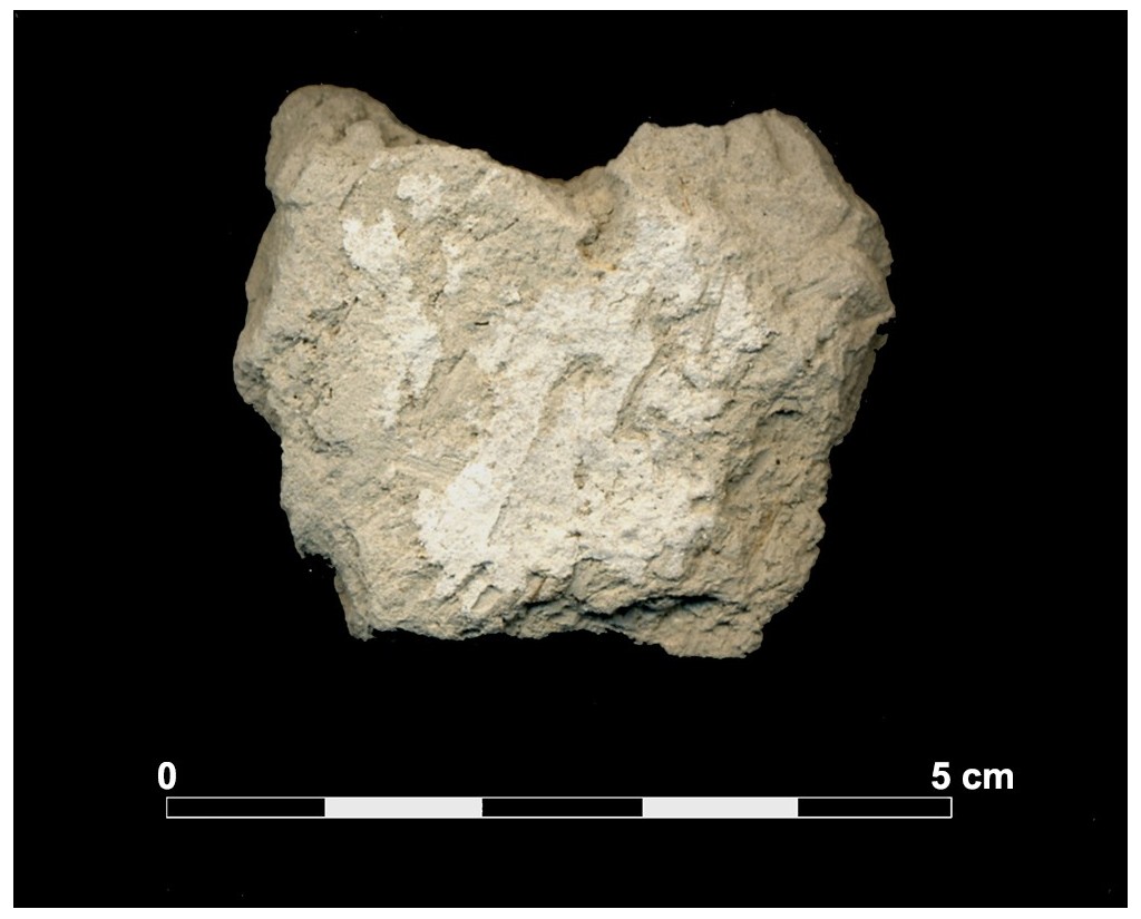

Covering 20010. Burial mound 20. Cemetery of Tutugi.
Dimensions
: 4 Centimeters
: 3 Centimeters
Materials
covering
Temporal
: Iberians, Iberian
: Late 5th ct. BC
Spatial
- : Cemetery of Tutugi
- : Galera, Granada, Spain
- : WGS84
Copyrights
Creative Commons - Attribution, Non-Commercial, No Derivatives (BY-NC-ND)
References
Rodríguez Ariza, Mª O. (2014): La necrópolis ibérica de Tútugi (2000-2012). CAAItextos. Universidad de Jaén, Jaén
Digital Resources
-
 Instituto Universitario de Investigación en Arqueología Ibérica
Creative Commons - Attribution, Non-Commercial, No Derivatives (BY-NC-ND)
Arquiberlab
http://creativecommons.org/licenses/by-nc-nd/3.0/
[ver más...] [ver menos] -
 Instituto Universitario de Investigación en Arqueología Ibérica
Creative Commons - Attribution, Non-Commercial, No Derivatives (BY-NC-ND)
Arquiberlab
http://creativecommons.org/licenses/by-nc-nd/3.0/
[ver más...] [ver menos] -

Creative Commons - Attribution, Non-Commercial, No Derivatives (BY-NC-ND)
Arquiberlab
http://creativecommons.org/licenses/by-nc-nd/3.0/
[ver más...] [ver menos] -

Creative Commons - Attribution, Non-Commercial, No Derivatives (BY-NC-ND)
Arquiberlab
http://creativecommons.org/licenses/by-nc-nd/3.0/
[ver más...] [ver menos]
Activities
University Research Center for Iberian Archaeology |
|
Spectroscopic analysis Mineral analysis of the outside white covering Non destructive. Surface cleaning. Sample pretreatment is not required. Direct measurement. Micro-raman Spectroscopy (MRS) Renishaw ‘in via’ Reflex Spectrometer coupled with a confocal Leica DM LM microscope (CICT, University of Jaén), equipped with a diode laser (785 nm, 300 mW), and a Peltier-cooled CCD detector, calibrated to the 520.5 cm-1 line of silicon. |